


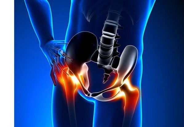
Hip joint arthrosis (coxarthrosis)- It is a chronic degenerative joint disease, which leads to deformation of bone tissue.With coxarthrosis, all components of the joint are involved in the pathological process: articular cartilage, bone structures adjacent to cartilage, synovial shell, ligaments, capsules and adjacent muscles.In the case of the disease, the articular cartilage is destroyed, bone and osteophyte micro-redinations appear (bone growth), and an inflammation of the muscular-ligamentous hip joint appears.
In the world, every fifth person complains of common problems with the nodes.This can also be the pain or restriction of movement in the joints, and a combination of these symptoms.The second ambulance vision falls in patients with bone-muscular disorders, while 66 % of cases are people under 65 years of age.According to the latest epidemiological research, the prevalence of knee joint arthrosis and hip among the adult population is 13 %.
Risk factors for the development of coxarthrosis:
- Genetic predisposition.A common cause of hip joint coxetrosis is congenital or acquired type of type II type of type.
- Old age.The potential cause of the spread of arthrosis in old age is a discrepancy between the harmful effect on the joint cartilage of the external environment and its ability to restore.
- floorWomen suffer from osteoarthritis more often than men.This is due to the effects of the influence of female sex hormones of estrogen on bone-mineral metabolism.However, the impact of the floor is unclear - according to some authors, unlike other joint damage, there are no sexual basis for coxartrosis: in men, hip joint arthritis is so often found in women.
- Excessive body weight.The relationship is confirmed between excessive body mass and the appearance of arthrosis.Excess adhesive tissue increases the harmful load on the cartilage.Moreover, adipose tissue produces pro -inflammatory enzymes that damage the cartilage tissue.
- Frequent development of bones and joints.In accordance with studies, 80 % of coxarthrosis, which occurs without any obvious reason, is associated with previously defects that are not diagnosed in the development of hip joint - dysplasia and subluxation.
- Heavy physical work.An excess load on the hip joints with certain types of physical work can lead to damage to the joints and the formation of arthrosis.At risk are agricultural workers, excavators and people of similar work specialties.
- Injuries.The risk of developing coxarthrosis increases after an injury to the hip joint.Moreover, both a damaged joint and both can be included in the process.
- Professional sports playing.Professional sport can provoke the emergence of coxarthrosis both due to excess load on the joints and due to damage.Potentially dangerous sports include heavy athletics, athletic jumps, parachute sports.
- Bones and common diseases- Rheumatoid arthritis, psoriatic arthritis, joint infections, avascular necrosis, gouty arthritis, etc.
- Endocrine- Hypothyroidism, hypoparathyroidism, acromegaly (damaged function of the anterial pituitary gland), diabetes, overweight.
If similar symptoms are detected, consult a doctor.Don't have it yourself -My - it's dangerous to your health!
Symptoms of hip joint arthrosis
The main symptoms of coxarthrosis include: pain, mobility restrictions and cramping on the joints, deforming them, functional shortening of the lower extremities and periodic swelling in the joints.
Pain of different intensity.The pain in the joints is initially irrelevant and arises for a short time.They appear or intensify during walking or with other physical exercises, for example, during meetings, tendencies and weight lifting.As the disease develops, the pain intensifies and even a long break does not cause relief.In addition, the pain appears with prolonged real estate and fastening the joint in one position.
Patients complain of so -called "starting" pains in the hip after sleeping joints, driving in a machine and other elongated real estate.Pain "starting" for coxarthrosis lasts no more than 30 minutes.Pain intensifies during hypothermia or in a stressful situation.They can be located in the area of the buttocks or groin, on the anterior or lateral surface of the thigh.With the spread of pain over the nerves of the lumbar plexus, it can be transmitted to the thigh away from the center of the body or knee.Sometimes the pain applies to the lumbosacral spine and the tail.
Restriction of joint mobility.Movements in the hip joint with coxartrosis are limited due to pain.At the same time, rotation (turns both inside and outside) and the behavior of the lower limbs (movement between the body) is worried more often, but may be limited (movement from the middle axis of the body), as well as bending and stretching.The inability to make passive movements in the fusion due to a pronounced pain syndrome causes a bias of the compensatory pelvis.The patient's walk changes, the buttocks climb back, the body deviates forward when transferring weight to the damaged side.With bilateral damage to patients with coxartrosis, a "duck walking" is formed.
With coxarthrosis occurs periodicallyEllingment in unionwhich can be invisible due to the muscle and fat layer.Also, the disease is characteristicCrystals in the joints during movement, their gradual deformity and functional shortening of the lower extremities.
Often, a fusion is affected by the disease, then the process applies to others.But sometimes arthritis affects some joints immediately and polyosostoarthritis occurs.Poliosteoartrosis is characteristic of the elderly or with an inherited predisposition and concomitant disease - bone disease, joints and endocrine disorders.
The pathogenesis of hip joint arthrosis
In the pathogenesis of hip joint arthrosis, an important role is played by mechanical damage (damage and microtraums due to increased physical exercise in the joint) and genetic, hormonal and metabolic factors.It is often not possible to find out which factor has affected the development of the disease in a particular patient, but often the disease develops after damage to tissue with mechanical damage.
Tissue damage stimulates the separation of cartilage tissue cells (chondrocytes), while the production of pro -inflammatory cytokines increases, which are normally present in the cartilage in only small quantities.Cytokines begin the inflammatory process, for example, under the influence of pro-inflammatory cytokine IL-1, enzymes that destroy the joint are distinguished.Also, under the influence of cytokines, the production of the TSOG-2 enzyme and other substances that have a toxic effect on the cartilage increases.
Synovites also play a major role in the development of coxarthrosis - inflammatory disease of the synovial shell of the joints or ligaments with fluid accumulation in the cavity.
A decrease in elasticity and articular cartilage strength associated with metabolic disorders leads to a decrease in its resistance to mechanical stress.With coxartrosis, all joints of the joints are involved in the pathological process, including a subcondrial bone.Due to the fact that large joints of the lower extremities constitute large joints of the body, they undergo significant mechanical stress, due to which the microwaves in the subcondral plaque and cartilage occur.As a result of the microvelomas, the subcondrial bone is compacted, which leads to regional growth of bone tissue - osteophytes.And this, in turn, stimulates further degradation of the articular cartilage.
In some cases, hip joint arthrosis is inherited.Inherited arthritis is supposedly polygenic inheritance - due to the action of many genes, each of which is poorly affected.The cause of some diseases is a mutation in genes that encode the articular cartilage macromolecules, which causes its fractures.The genes responsible for separating the chilocytes can also suffer.Moreover, metabolic disorders are inherited, such as pyrophosphate arthropathy - a disease in which calcium pyrophosphate crystals accumulate in articular cartilage and synovial fluid.
Classification and stages of development of hip joint arthrosis
Depending on the causes of the disease, coxarthrosis is divided into two main forms: primary (idiopathic) and secondary (derived from or due to other diseases).
Primary coxartrosis:
- Localized (only hip joints affect):
- biased;
- bilateral.
- Generalized (polyosteoarthrosis) with a lesion of at least three common groups (for example, hip, knee and small joints of brushes or legs).
Secondary arthrosis:
- Post -traumatic:
- Acute - as a result of acute damage;
- Chronic - due to the classes of some sports or as a result of professional activity.
- Metabolic diseases (okonosis, hemochromatosis, Wilson's disease, Gaucher disease).
- Congenital pathologies and development defects (congenital hip coupling dysplasia, disease, sliding of the femur, hypermobility syndrome, lower extremity, scoliosis, bone dysplasia).
- Endocrine pathologies (acromegaly, hypothyroidism, diabetes mellitus, hyperparathyroidism, overweight).
- Calcium salts (pyrophosphate arthropathy, calcifying tendonitis).
- Bone and joint diseases (rheumatoid arthritis, psoriatic arthritis, pehetic disease, avascular necrosis, infections).
According to clinical manifestations, the following forms of coxarthrosis are distinguished:
- Few symptoms.
- Manifest, manifested by bright clinical symptoms:
- Quickly progressive, in which symptoms develop in the first four years from the onset of the disease;
- Slowly progressive - important clinical symptoms appear after five years of the course of the disease.
In accordance with figure X -Ray, two types of hip joint arthrosis can be identified:
- Hypertrophic - with signs of increased compensation reply (lesions are replaced by a new tissue, for example, osteophytes appear);
- Atrophic (decreased tissue volume).
The stages of the disease can be radiologically and clinically determined.To determine the radiological phase of the arthrosis of the hip joint, the classification of Kellgren and Lawrence (1957) is most often used.
Stages of arthrosis in radiological classification
| Scene | Signs |
|---|---|
| 0 | No signs of arthrosis in images x -ra |
| 1 | The common gap has not changed, the only regional osteophytes are visualized |
| 2 | The common gap has not changed, the important regional osteophytes are visualized |
| 3 | The height of the common gap is moderately reduced, the important regional osteophytes are visualized |
| 4 | The height of the joint gap is significantly reduced, important regional osteophytes and subcondral osteosclerosis are visualized (bone tissue compression below the cartilage surface with the cartilage structure) |
To determine the clinical phase of the disease, classification (1961) is used, which uses both clinical signs and visualization criteria.
The clinical stages of arthrosis
| Scene | Signs |
|---|---|
| 0 | The articular gap is narrowed without hesitation and uneven, the edges of the articular cracks are slightly pronounced (initial osteophytes), a small restriction of movements is marked |
| 1 | The articular gap is significantly narrowed (50-60 %), significant osteophytes, subcondrial osteochosclerosis and cystic enlightenment in bone epithet;The clinic prevails by restriction of mobility to the joints, a rough crisis during movements, unnecessary or moderate muscle atrophy |
| 2 | deformity, stiffness of the joint;The articular gap is narrowed by more than 60-70 % of the rate or widely missing osteophytes, subconders, "articular mice" are visualized bone, cartilage or mixed pathological formations located in the joint cavity |
Hip joint arthrosis complications
With coxarthrosis, all complications are associated precisely with pathological changes in the joints.
The course of coxarthrosis can be complicated by local inflammatory processes:
- Bursite - inflammation of synovial bags in the joints;
- Tendovaginitis - inflammation of the inner shell of the vagina of muscle tendons;
- Nerve-pins syndrome of nerve due to the formation of large osteophytes or with joint deformity.
With the progress of coxarthrosis and its transition to clinical stages II and III, pain in the movement of the joint occurs, and over time, joint ankylosis (fibrous, bone or cartilage) occurs, accompanied by its complete immobility.
Significant common deformation can lead toFractures or aseptic bone necrosis.For coxartrosis, the aseptic necrosis of the femur's head is the most frightening complication.
With pronounced coxarthrosis, may occursubluxation and shift of joiningas well as penetration of the femur's head into the pelvic cavity.The displacements and subluxation of the hip joint lead to pain (at first acute, then dull and pain), intensifying during walking and other physical exercises, as well as in the deformation of the joint, lame, and sometimes to cut the affected limb.
Despite the lack of systemic manifestations of arthrosis itself, in modern clinical practice, more attention is paid to it related diseases.These are such pathological conditions that exist or arise against the backdrop of current disease.With regard to inflammatory reactions arising during arthrosis, the formation of atherosclerotic plaques in the inner walls of the vessels has improved, which increases the riskCardiovascular disease.A decrease in physical activity due to pain and restriction of joint mobility leads toObesity, depression and deterioration in the quality of life.With prolonged use of non -steroidal anti -inflammatory drugs,The upper gastrointestinal sections are affected,And tooThe risk of cardiovascular pathologies and kidney disease increases.
Diagnosis of hip joint arthrosis
The diagnosis of "coxarthrosis" is made on the basis of clinical manifestations and radiological examination.There are no characteristic laboratory signs for the diagnosis of arthrosis.
Among the clinical manifestationsThe main one for the diagnosis of arthrosis of the hip joint is its pain and character.Pain for hip joint arthrosis occurs and increases gradually over several years (sometimes several months with a rapid progressive form).Pain occurs or increases during physical exercise or in a standing position.If the patient begins to feel pain only, then inflammation (synovitis) joined.The statement is recorded up to 30 minutes in the morning and with prolonged immobility.
The restriction of joint mobility is gradually increasing, this applies to active and passive movements.With the development of the disease, the joints are deformed, the functional shortening of the limb length can occur.
In an examination of the physicalThere is a restriction of joint mobility, their deformity, limb shortening, pain in the joint palpation and a large femur rotation, muscle atrophy.
Laboratory methodsFor the diagnosis of hip joint arthrosis is not required.However, they can be used for the differential diagnosis of coxarthrosis with arthritis (rheumatoid and chronic), as arthrosis has no inflammatory changes in the overall blood test and rheumatoid factor, and uric acid levels have not increased.Moreover, using laboratory tests, contraindications are detected for medication treatment methods.
Instrumental methodsFor the diagnosis of hip joint arthrosis:
- Radiograph- This is the main method of diagnosing arthrosis of the hip joints.Radiography determines the characteristic changes of coksartrosis: narrowing of the common gap, osteophyte, erosion and cartilage ulceration, subcondular cysts and osteosclerosis.X -Ray examination is a classic method for diagnosing coxarthrosis, and radiological signs underline the classification of coxarthrosis.However, currently, other methods of joining visualization are increasingly used, such as ultrasound and magnetic resonance imaging.
- Ultrasound examination (ultrasound) -The advantage of ultrasound is in the absence of a radial load on the body.
- Magnetic Resonance Tomography (MRI)- Compared to other methods, it allows you to visualize the damage to the joints more clearly.
- arthroscopy-It allows you to identify articular cartilage damage: from chondromation areas (softening of articular cartilage) with a diameter of less than 10 mm to deep cracks penetrating up to the subcondition bone and the formation of deep ulcers.Surface and medium acks and surface erosion can also be visualized.
Identifying coksartrosis usually does not present particular difficulties, but when evaluating a specific clinical situation, it is necessary to remember the possible secondary origin of hip arthrosis (as complications of other diseases, for example, with endocrine disorders).
Hip joint arthrosis treatment
The treatment of hip joint arthrosis can be conservative (medicine and not united) or operational.Conservative treatment is used in stages 1-2 of the disease, surgical-in 3 phases.Surgical treatment can be recommended in 2 stages with persistent pain and lack of response to conservative therapy.
Conservative therapy goals:
- improve quality of life - reduce pain and increase joint mobility;
- Stop or slow down the development of the disease.
Non -Drug treatment methods include:
- discharge of the hip joint (lowering body weight, creating additional support and transfer of part of body weight to cane or crutches);
- Physical education of physiotherapy;
- Methods of physiotherapeutic treatment.
Treatment of coxarthrosis begins with non -drug methods, an important role is given to physiotherapy exercises.With severe pain, the patient should use support.With a pronounced disease and the presence of contraindications to endoprosthetics, support should be used for life.
Cuxartrosis medicinal therapyIncludes medicines that reduce the symptoms of the disease.These are analgesics as well as medicines from the group of non -steroidal anti -inflammatory drugs (NSAIDs).NSAID are divided into non -election and selective.
Analgesics and NSAIDs for hip joint arthrosis are used for a short time to relieve pain and inflammation.Currently, there is no proven advantage of a non -steroidal anti -inflammatory agent over another, so the choice of a particular medicine depends on the side effects and a specific clinical situation caused by it.
It should be remembered that NSAIDs have a number of side effects.When you take them, the mucous membrane of the stomach and duodenum is affected, as a result of which ulcers and bleeding are possible.A number of NSAIDs have a toxic effect on the liver and kidneys.Moreover, NSAIDs disrupt platelet accumulation, and, as a result, the patient is terminated by thrombosis and there is a tendency for bleeding.NSAIDs with prolonged use suppress the processes of hematopoiesis and can cause aplastic and agranulocytosis anemia.Reception of selective NSAIDs causes significantly less complications.
Ointments and gel used in the country cause fewer side effects than oral products.For the treatment of arthrosis, medicines with heat and low pain are used.They can contain nicotinic acid esters, salicylate, propeller, bee venom.Also, NSAIDs have a good effect.
In the absence of the effect of analgesics and NSAIDs or if it is impossible to choose the optimal dose of the drug, the painkillers of central action may be prescribed short -term.
In the case of inflammation, intra -articular corticosteroids are used.Corticosteroids are used no more than 2-3 times a year, as the most common use can lead to cartilage degeneration.
Slowly drug drugs weaken the symptoms of the disease include chondroprotectors, inappropriate compounds of avocado or soybean, hyaluronic acid.These medicines are included in the recommendations of European antiraematic bonding for the treatment of hip joint arthrosis.Preparations reduce pain and improve joint mobility.
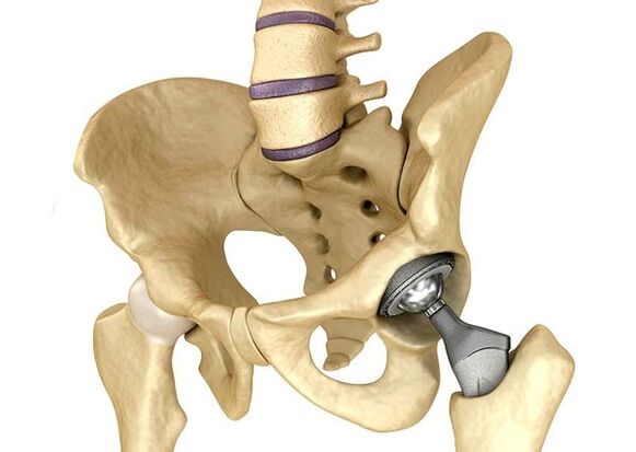
Endoprostetics of hip jointsIt is used in severe cases of phase III, when the pain syndrome cannot be eliminated, and the motility of the joint is significantly limited.Hip joint prosthetics leads to a decrease in pain syndrome, an improvement in the functional state of the joint and the quality of life of the patient.The effect persists for 10-15 years, after which a second operation may be required.During surgery, the hip node is replaced by artificial imitation of ceramics, metals (most commonly used titanium prostheses) or polymer.
Forecast.PREVENTION
The prognosis of hip joint arthrosis about the patient's life is favorable, but the disease often leads to disability.According to the World Health Organization, 80 % of elderly patients with coxarthrosis have a violation of mobility, and 25 % cannot make daily issues.In this regard, the main prevention of arthrosis of the hip joints is important.
Prevention measures:
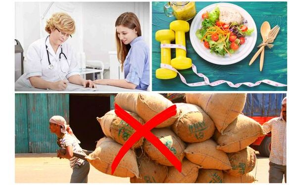
- Lower the body weight.It is necessary to adjust the food in order to reduce the weight and load on the joint.Moreover, a decrease in adipose tissue volume reduces the amount of inflammation mediators it released.
- Avoid heavy physical and sporting overloads.Physical overloads are often the cause of arthrosis of the hip joints, while moderate physical activity, on the contrary, improves the condition of the articular cartilage, maintains its normal movement and reduces the load on other joints.
- Correct the underlying disease.If the patient is detected in diseases that can lead to secondary coxartrosis (endocrine, rheumatic and others), the underlying disease is necessary.Normalizing the hormonal background and achieving continuous remission of rheumatic diseases is also the main prevention of arthrosis, and allows you to slow its development.
- Run a healthy lifestyle.A balanced diet with sufficient content of plant and animal proteins, unsaturated fatty acids and limiting simple carbohydrates, as well as moderate physical activity, avoid the appearance of coxarthrosis even in the presence of risk factors.
Currently, prevention of hip union diseases is mandatory in neonatology and pediatrics.Over time, the regulated congenital dysplasia of the hip joint significantly reduces the risk of coxarthrosis in adulthood.